微信登录
微信登录 收藏本站
收藏本站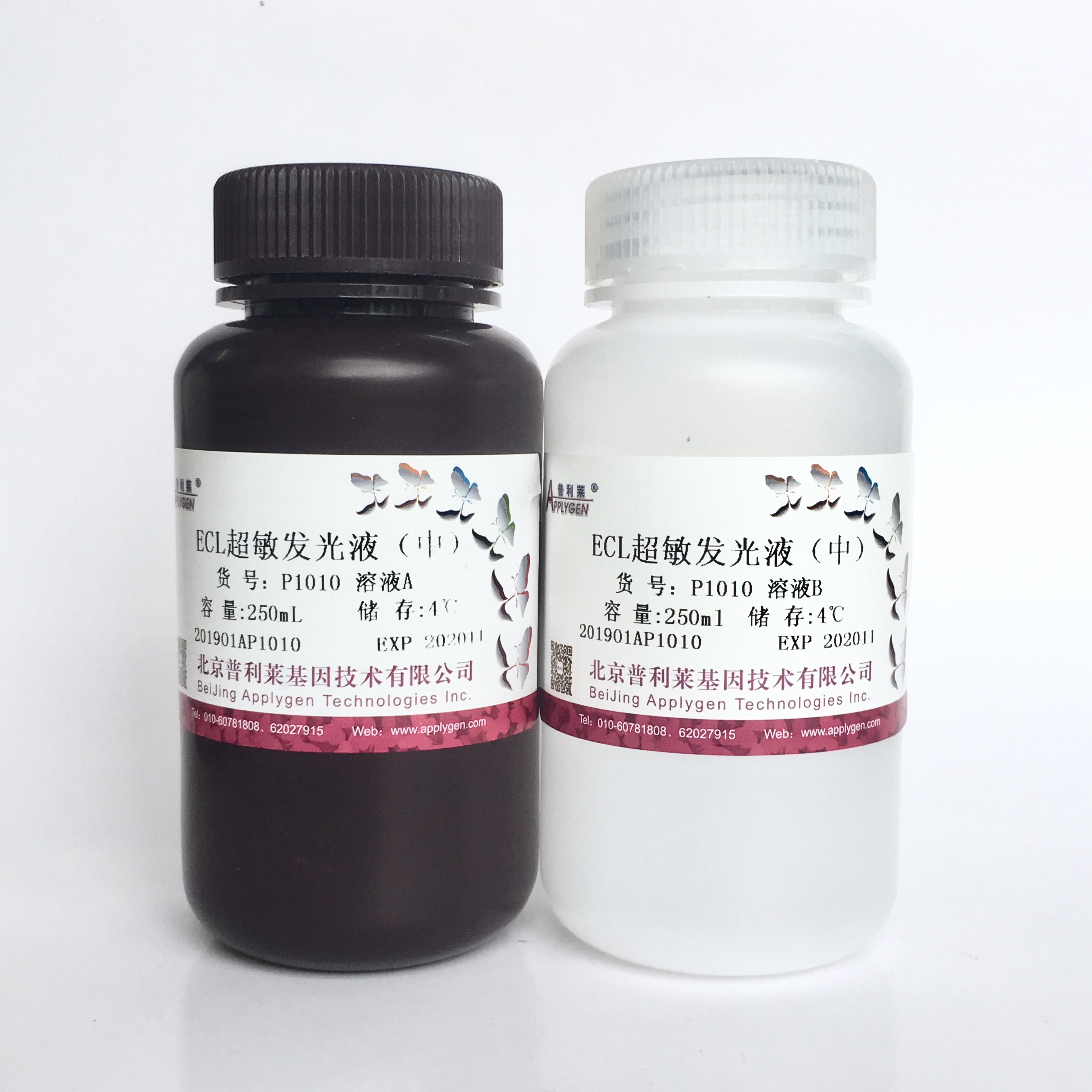
ECL超敏发光液(中) P1010提供OEM定制服务,大包装更优惠!

- 产品详情
- 说明书下载
- 引用文献
描述:
ECL超敏发光液 (中)用于直接或间接检测与辣根过氧化物酶HRP关联的蛋白或核酸底物,适用于大多数蛋白的ECL化学发光。
特点:
1、 灵敏度高,背景干净
2、 发光迅速,节省抗体
3、 操作简便,适用于机器扫描和胶片曝光
规格:25mL 100mL 250mL 500mL
储存: 4 ℃避光储存1年
效果展示:

使用方法:
1. 常规电泳、转膜、HRP标记抗体或核酸探针孵育、洗膜。注意用HRP标记lgG或用一抗-链亲和素-生物素-HRP夹法。核酸杂交膜用HRP标记探针杂交,洗膜。
2. 洗涤膜上的HRP标记二抗时,配制新鲜发光工作液:分别取等体积的溶液A和B混合,放置使之升到室温否则会减弱荧光强度。建议立即使用工作液,室温放置数小时后仍可使用但灵敏度略有降低。
3. 用镊子取出膜,搭在滤纸上沥干洗液但勿使膜完全干燥。将膜完全浸入并与发光工作液(0.125mL发光工作液/cm2膜)充分接触。准备立即使用全自动化学发光成像系统扫描。孵育时间过长不会增加灵敏度,有时还会导致曝光条带异常。发光过程的本质是酶促反应,使用过少的发光工作液不利于反应进行,也会导致膜上条带曝光不均,明显降低灵敏度。为达节约目的可将膜剪小但勿降低发光液用量。
4. 用镊子夹起膜,搭在滤纸上沥干发光工作液。但勿洗去发光液。
5. 全自动化学发光成像系统扫描或胶片曝光。
发表文章时,可参考以下格式:
The protein bands were visualized by enhanced chemiluminescence detection reagents (Applygen Technologies Inc., Beijing, China)
目前,使用我公司线粒体/胞浆蛋白制备试剂盒发表的SCI英文论文已有多百篇,以下供参考:
1、Shangguan W J, Li H, Zhang Y H. Induction of G2/M phase cell cycle arrest and apoptosis by ginsenoside Rf in human osteosarcoma MG 63 cells through the mitochondrial pathway[J]. Oncology reports, 2014, 31(1): 305-313.
2、Zhou X, Zhao Y, Fang Y, et al. Hes1 is upregulated by ischemic postconditioning and contributes to cardioprotection[J]. Cell biochemistry and function, 2014, 32(8): 730-736.
3、Zhang P, Pan H, Wang J, et al. Telomerase activity-independent function of telomerase reverse transcriptase is involved in acrylamide-induced neuron damage[J]. Biotechnic & Histochemistry, 2014, 89(5): 327-335.
4、Zhou X L, Wan L, Xu Q R, et al. Notch signaling activation contributes to cardioprotection provided by ischemic preconditioning and postconditioning[J]. J Transl Med, 2013, 11: 251.
5、Zhang F, Zhang L, Sun L, et al. Effects of Fluid Shear Stress on Expression of Smac/DIABLO in Human Umbilical Vein Endothelial Cells[J]. Current Therapeutic Research, 2013, 74: 36-40.
6、Zhao G, Ma H, Shen X, et al. Role of glycogen synthase kinase 3β in protective effect of propofol against hepatic ischemia–reperfusion injury[J]. Journal of Surgical Research, 2013, 185(1): 388-398.
7、Sun L L, Zhang L, Meng X L, et al. Effects of fluid shear stress on the expression of Omi/HtrA2 in human umbilical vein endothelial cells[J]. Molecular medicine reports, 2013, 7(1): 110-114.
8、Sun L Q, Zhao J, Zhang T T, et al. Protective effects of Salvianolic acid B on Schwann cells apoptosis induced by high glucose[J]. Neurochemical research, 2012, 37(5): 996-1010.
9、Zhou Q, Li Y, Jin J, et al. Lx2-32c, a novel taxane derivative, exerts anti-resistance activity by initiating intrinsic apoptosis pathway in vitro and inhibits the growth of resistant tumor in vivo[J]. Biological and Pharmaceutical Bulletin, 2012, 35(12): 2170-2179.
10、Li W, Zhang J, An W. The conserved CXXC motif of hepatic stimulator substance is essential for its role in mitochondrial protection in H 2 O 2-induced cell apoptosis[J]. FEBS letters, 2010, 584(18): 3929-3935.
11、Fang N X, Yao Y T, Shi C X, et al. Attenuation of ischemia–reperfusion injury by sevoflurane postconditioning involves protein kinase B and glycogen synthase kinase 3 beta activation in isolated rat hearts[J]. Molecular biology reports, 2010, 37(8): 3763-3769.
12、Zhao C Q, Zhang Y H, Jiang S D, et al. Both endoplasmic reticulum and mitochondria are involved in disc cell apoptosis and intervertebral disc degeneration in rats[J]. Age, 2010, 32(2): 161-177
13、Yin Q, Jin P, Liu X, et al. SDF-1α inhibits hypoxia and serum deprivation-induced apoptosis in mesenchymal stem cells through PI3K/Akt and ERK1/2 signaling pathways[J]. Molecular biology reports, 2011, 38(1): 9-16.
相关产品